Spectral Shift
The novel method for ultra-fast and precise detection of molecular interactions What can you do with it?How does it work?Get binding affinities as close as possible to reality

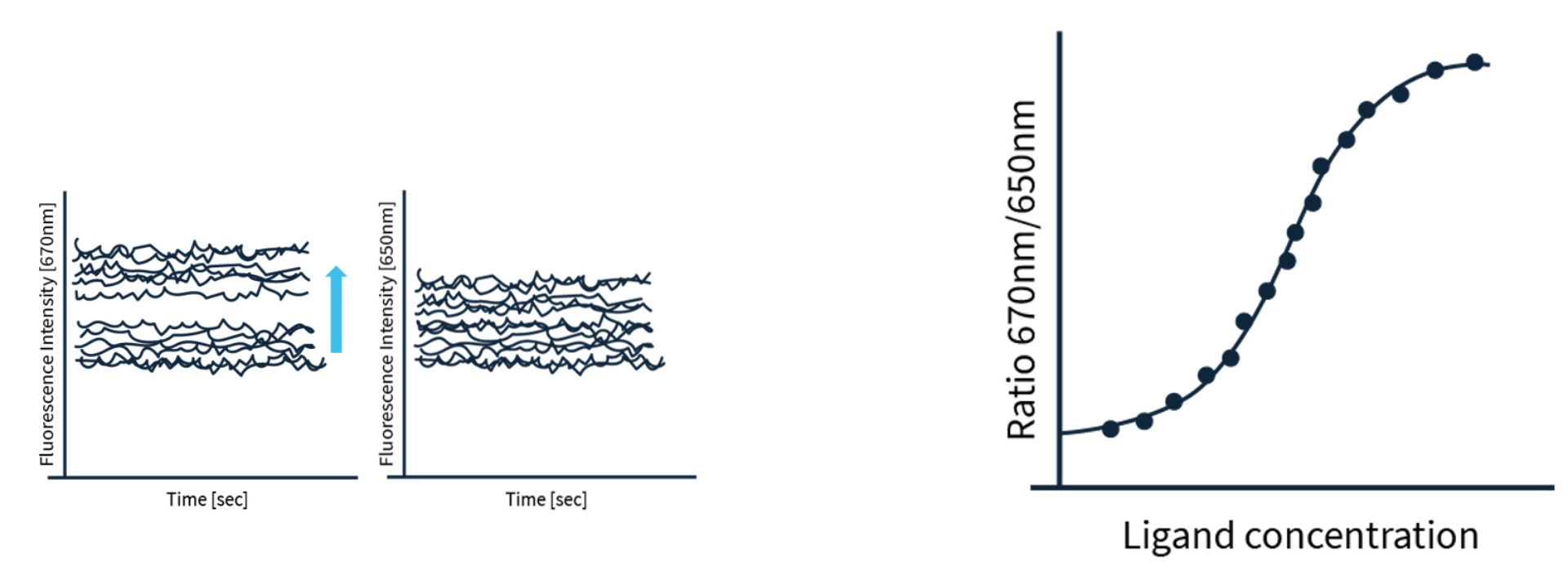
Spectral Shift is a novel tool for detection and analysis of molecular interactions and KD-determination. It works fully in solution and isothermally at room temperature by applying an extremely sensitive (sub-nm) fluorescence shift detection. All that while using practically no sample at all (down pg of protein per experiment).
2bind’s Spectral Shift assays can be scaled from just a single interaction up to 10.000’s of interactions.
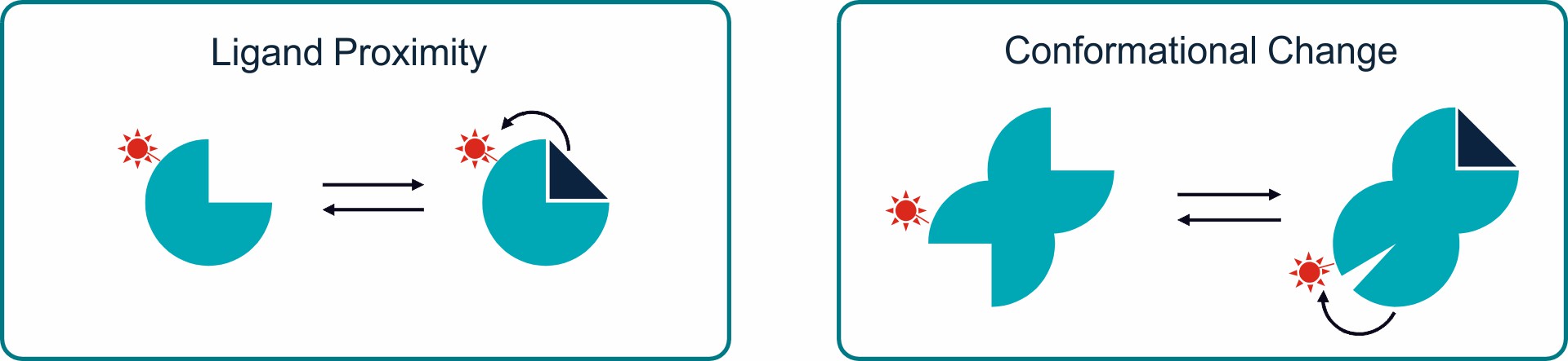
Spectral Shift works with almost all classes of target molecules as long as they can be made fluorescent. The variety of ligand molecules is even greater – from molecular fragments up to virus-like particles. The fluorescent target molecule is kept at a constant concentration and the non-fluorescent ligand molecule is titrated across a large concentration range.
Affinity
LIGAND BINDING
KD
Equilibrium dissociation constant. Can be obtained by kinetic or classical equilibrium binding analysis. Provides information about the strength but not the dynamics of an interaction.
ΔH
Binding enthalpy. KD values at different temperatures can be used to obtain the binding enthalpy of an interaction via vant-Hoff-plots.
Ligand scouting
Fast screening for yes/no binding answers.
Other assay types
COMPETITION ASSAYS
Determination of competition of several ligands for a selected binding site on a target.
COMPLEX INHIBITION ASSAYS
Determination of binding site blocking by a ligand and prevention of binding of a natural interaction partner.
STOICHIOMETRY ASSAYS
Determination of equilibrium binding stoichiometry of target-ligand complex formation.
What Spectral Shift can tell
Single-dose screen
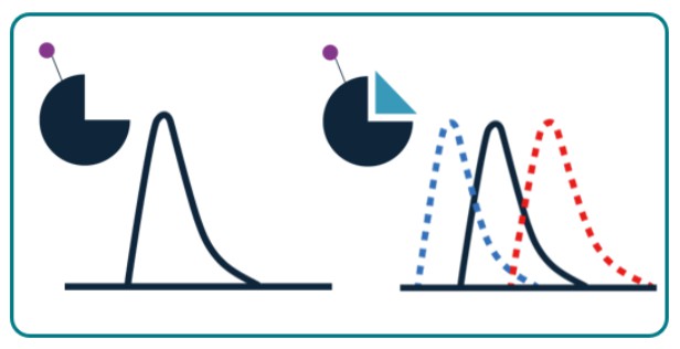
Spectral Shift will tell you whether a target and ligand generally interact from a single-dose experiment.
Steady-State Affinity
Spectral Shift will tell you the precise and true steady-state affinity (dissociation constant KD) of your interaction.
Protein Aggregation
Spectral Shift will tell you whether your protein samples aggregate or if aggregation is induced by ligand binding.
Ligand competition
Spectral Shift will tell you competitive effects between multiple binders of your targets.
- Drug discovery (compound-based and fragment-based)
- Drug discovery (protein-targeted and RNA-targeted)
- Protein characterization
- Additive screening
- Enzyme-inhibitor screening
- Drug discovery (compound-based and fragment-based)
- Drug discovery (protein-targeted and RNA-targeted)
- Protein characterization
- Additive screening
- Enzyme-inhibitor screening
- Protein and enzyme quality control
- Buffer screening and optimization
- Protein storage optimization
- Ligand-induced aggregation testing
- Complex binding analysis
- Mode-of-action analysis
- Competitor screening
- PPI-disruption analysis
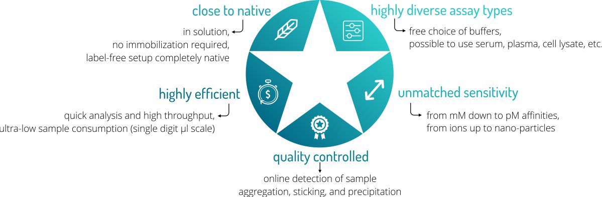
Advantages of Spectral Shift
2bind Spectral Shift Services
2bind offers a variety of services using the Spectral Shift technology. Benefit from fast and precise analysis of molecular interactions.
Enjoy minimal sample consumption (just nM concentrations and µL volumes) and robust analysis methods. The 2bind Spectral Shiftservices can be combined as you choose for all questions in drug discovery, antibody development, protein biophysics and analysis, as well as aptamer characterization.
Spectral Shift Technology and FAQs
Overview
Technology
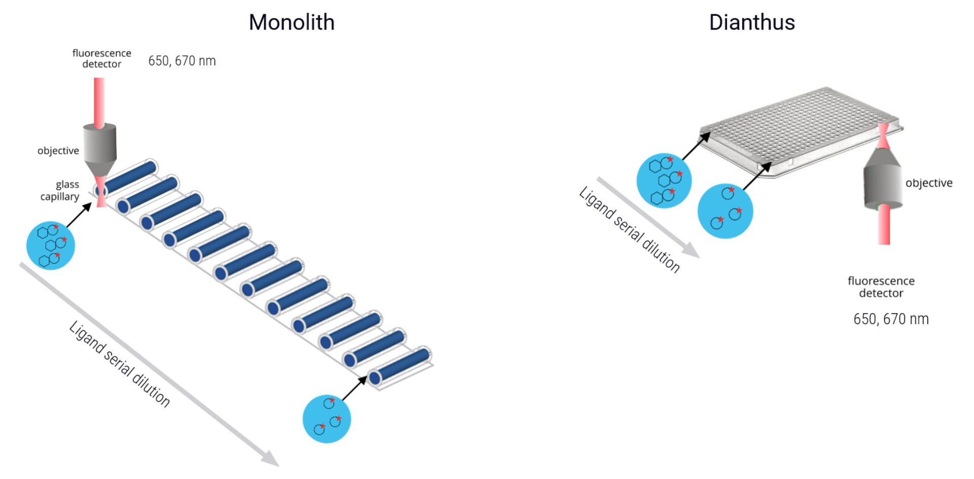
MST measurements are possible either in a glass capillary setup (Monolith-series instruments, NT.Automated) or in a plate-based format (Dianthus). In both setups, an infrared laser is used to generate a precise temperature gradient while an LED is used for the excitation of fluorescent molecules inside the glass capillaries or the plate wells. The capillaries or wells each contain a mix of fluorescent binding partner (usually called the “target” and non-fluorescent binding partner, which is titrated in a dilution series (usually called the “ligand”).
The sample fluorescence in each capillary or well is measured over time. After a certain time, the infrared laser is switched on and the sample is heated. Such resulting MST traces are recorded for each ligand concentration and plotted against time. The calculated Fnorm values from the MST traces (fluorescence in region F1 divided by fluorescence in region F0) are dose-dependent and can be well described by the law of mass action. A plot of Fnorm against ligand concentration then returns the dissociation constant KD of the interaction.
MST allows for the monitoring of either fluorescently-labeled molecules or intrinsically fluorescent molecule (such as proteins; the latter would be a truly label-free measurement). MST measurements are possible in any kind of buffers, even in serum, plasma, cell lysate, urine, mucus, or other environmental matrices. Data generation is fast and precise and the data output is comparable to other biophysical methods.
Typical applications
Typical applications of MicroScale Thermophoresis include:
- Hight-throughput target-ligand interaction screenings
- Steady-state binding affinity assay
- Steady-state binding affinity assay in biological liquids
- Sandwich assays (1 target, 2 ligands)
- Competition assays
- Binding affinity assays with multiple binding partners
For more information, please visit our Application Database. The different service areas can be found under MST Services.
Compatible Fluorescent Dyes
Spectral Shift relies on measuring the fluorescence of the studied molecules. Thus, one of the binding partners has to be fluorescent. Different fluorescent methods and/or labelings are possible. Most commonly, the labels listed in the table below are used. 2bind performs the labeling of your target protein for you with all possible dyes.
In the case of DNA or RNA target molecules direct covalent linkage of fluorophores (e.g. Cy5) has proven well.
The following table gives an overview over the most commonly used fluorophores in Spectral Shift. Please keep in mind that, in principle, every fluorophore can be used as long as its excitation and emission wavelength ranges match the ones of the fluorophores listed here.
| Fluorophore | Excitation (nm) | Emission (nm) |
|---|---|---|
| Cy5 | 649 | 670 |
| NT-647 (RED) | 650 | 680 |
| Alexa647 | 652 | 668 |
| NT-RED 2nd generation | 650 | 670 |
Advantages
Spectral Shift offers a number of great advantages over other biophysical methods for determining the affinity of a molecular interaction.
- Low sample consumption → minimum of only 6 µl is required per sample
- Free choice of assay buffers → also biological liquids possible such as serum or cell lysate
- Very short analysis time → short analysis time enables high throughput
- Real-time quality controls → online aggregation, precipitation, and sticking controls
- Wide temperature range → analysis possible from 20°C to 45°C
- No immobilization required → measurement is done truly in solution
- Wide concentration range → affinities can be analyzed in the pM-mM range
- Wide molecule size range → from 100 Da to 1 MDa
For a comparison of Spectral Shift with other biophysical techniques, please refer to our Technology Comparison Guide.
FAQ – General
What kinds of molecular interactions can I measure?
You can measure bi-molecular interactions between any kind of molecule: protein, RNA, DNA, small molecule compounds, lipids, carbohydrates (See figure on top of this page).
Is it possible to analyze interactions with aptamers?
Yes. Analyzing molecular interactions involving aptamers or other DNA- or RNA-molecules is no problem. Usually, Spectral Shift measurements with aptamers are straight-forwards, because these DNA molecules can be synthesized directly with fluorescent labels at their 5′ or 3′ end. Most commonly, Cy5 or Alexa647 are used as aptamer or DNA fluorophores.
More information on this topic is available in our Aptamer Services.
What information do I get from an Spectral Shift measurement?
Spectral Shift is not only able to determine the affinity and binding strength of a molecular interaction, but also allows for assessing other physical parameters such as stoichiometry, aggregation, precipitation, enthalpy (van’t Hoff plot), slow enzyme kinetics, and oligomerization.
For specialized kinetics measurements, consider Biolayer Interferometry. If you are interested in the thermodynamics of a molecular interaction, Isothermal Titration Calorimetry is an option. For an in-depth analysis of protein aggregation, protein stability and protein unfolding, take a look at the nanoDSF technique.
What type of fluorescent dyes can I use?
Our Spectral Shift instruments detect red fluorescent dyes as well as substrates with a wide variety of chemically attached fluorophores. Please see above (“Compatible Fluorescent Dyes”) for more information on the specific dyes that can be used.
Is it possible to measure without labelling a molecule?
No, in contrast to MicroScale Thermophoresis (MST), which also offers a “label-free” measurement setup (which relies on tryptophan fluorescence instead of an external fluorescent dye), Spectral Shift always required the labeling of the target molecules with a “red” fluorescent dye (see above for compatible dyes).
Can I measure binding kinetics with Spectral Shift?
Spectral Shift measures equilibrium binding constants (steady-state affinity). Typically, samples are inserted in the instrument after the binding reaction reaches chemical equilibrium. Measurement of kinetics is possible if the equilibrium is not reached quickly. On average, the kinetics of reactions that take more than 20 minutes can be evaluated.
An in-depth kinetics analysis of molecular interactions is possible with Biolayer Interferometry.
Can I measure thermodynamics with Spectral Shift?
Spectral Shift measures equilibrium binding constants (steady-state affinity) at a given temperature. By performing such experiments at different temperatures, the binding enthalpy can be derived from van’t Hoff analysis.
An in-depth thermodynamics analysis of a molecular interaction is possible with Isothermal Titration Calorimetry.
FAQ – Samples
Can I check for the quality of my samples?
Yes! Direct intrinsic quality controls available in the Spectral Shift setup allow for detecting protein aggregation as well as adhesion and sticking effects of the assay samples to the glass capillaries.
An in-depth analysis of the stability and aggregation behavior of your protein samples can be done with nanoDSF.
Is it possible to measure the binding of small molecules?
Yes, we are able to measure molecular interactions involving small molecules with the same high sensitivity and quality as for protein-protein, protein-DNA or protein-vesicle interactions. For more information on Spectral Shift and small molecule please have a look at our Drug Discovery Services.
What are the required concentrations for small molecules?
Dependent on the fluorophore (fluorescent label or intrinsic tryptophan fluorescence), between 1 – 100 nM of the labeled target molecule can be used. This target is titrated with the unlabeled small molecule compound in the range of ± factor 10 of the expected affinity (dissociation constant). For standard applications, 6 – 10 µl of sample material are used per data-point.
My protein is only stable at high ionic strength. Can I still test for binding with Spectral Shift ?
Yes, in general it is possible to work with the ionic strength that is best suited for your molecules. Most of the time, a buffer change is not required. For example, we have already successfully measured molecular interactions in a solution with a sodium chloride concentration of 1.5 M.
If you are not sure whether your sample protein really requires high ionic strength buffers, it is possible to investigate protein stability, thermal unfolding, and its aggregation behavior using nanoDSF. We offer a variety of Services in this respect.
I want to measure binding to a Mega-Dalton (MDa) molecule. Is this possible?
Yes! We have measured protein binding to the 70S ribosome unit as well as the binding affinity to liposomes with a size of more than 100 nm.
FAQ – Assay Conditions
Are there any limitations to the buffers or additives I can use?
No. You can use practically any buffer and any additive as long as it does not negatively affect the stability or homogeneity of your sample. Additives that affect the viscosity of the sample solution should be kept at a constant concentration.
A great way to check the tolerance of your protein samples for a vast number of additives is nanoDSF, a method that analyzes the thermal stability and unfolding of proteins. This technique can be used for buffer and additive screenings.
Can I measure in biological liquids like serum or lysates?
Yes. We are able to analyze the affinity of a molecular interaction also in complex biological liquids like crude cell extract, serum, plasma, urine, sea water, etc. You don’t have to pass on sensitivity or precision when analyzing these types of samples.
If you are interested in the stabilty of your protein sample, this can be determined using our nanoDSF technology.